Project Overview

Research Projects

Collage of microscopy images and graphic representations of cellular processes; the labels read “M. marinum”, “D. discoideum”, “exocytosis”, “ejection”, “cell death”, ‘PI4P’, “OSBP8”.
© Caroline Barisch

P1 - Caroline Barisch : Remodelling and exploitation of the host lipid trafficking machinery by pathogenic mycobacteria

We exploit Dictyostelium infected with mycobacteria to investigate how pathogens reprogram metabolic lipid flows and impact the functional plasticity of the mycobacteria-containing vacuole membrane composition.

Mikroskopische Aufnahme eines Gewebes, in dem einige Bereiche grün und andere violett gefärbt sind.
© Tim Koopmans

P2 - Kerstin Bartscherer : Formation, composition and role of extracellular vesicles in the ischemic heart

This project takes advantage of an ischemia model of highly regenerative spiny mice (Acomys) to ask how extracellular vesicles are formed in response to tissue damage and how they affect cardiac repair.

Several microscopy images with orange flashes, plus graphic representation of objects including a microscope; labels read “GFPA”, “GFPB”, “GFPC” and “Lipid handling”, as well as “Mito”, “Golgi”, ‘PO’ and “Organelle”.
© Maria Bohnert

P3 - Maria Bohnert : Plasticity of cellular lipid handling machineries in metabolic adaptation

We use microscopy-based screens to identify principles controlling the functional plasticity of lipid metabolism and inter-organelle lipid exchange, with a primary focus on sphingolipids, sterols, and storage lipids.

Collage of microscopy images and graphic representations of cellular processes; the labels include “Survival”, “Cytokines“ and ”Death".
© Katia Cosentino

Project P5 - Katia Cosentino : Plasticity of GSDM pores for the control of pyroptotic cell death

We analyze the role of the membrane environment on regulatory mechanisms of Gasdermin recruitment and pore formation at the plasma membrane in pyroptosis, an inflammatory form of regulated cell death.

Collage of microscopy images, models of molecules and graphical representations of experiments. The caption reads, among other things, “L-serine + palmityol-CoA --> 3-ketosphinganine --> sphingolipids”.
© Florian Fröhlich

Project P6 - Florian Fröhlich : Plasticity of the serine palmitoyl transferase complex during adaption of sphingolipid metabolism

We determine the function and regulation of the key enzyme in sphingolipid metabolism, the serine palmitoyltransferase (SPT) enzyme complex to understand metabolic sphingolipid fluxes.

Collage of microscopy images, a graph and a molecular structure.
© Ayelén González Montoro

Project P7 - Ayelén González Montoro : Dissecting the interplay between vacuolar contact site plasticity and sphingolipid homeostasis

We study the contact sites of the vacuolar membrane formed by the protein Cvm1, as a model to understand how contact site plasticity can determine membrane homeostasis.

Collage of microscopy images and graphic representations of cellular processes.
© Michael Hensel

Project P8 - Michael Hensel : Dynamic manipulation of functional plasticity of the host cell endosomal system by an intracellular pathogen

We explore how Salmonella infection and secretion of its effectors reprograms the mammalian endosomal membrane system to promote replication in its intracellular niche.

Collage of microscopy images, graphic representations of cellular processes and an X-ray image of a skull.
© Joost Holthuis; skull: Pekkinen et al. 2019, JCI Insight, https://creativecommons.org/licenses/by/4.0/

Project P9 - Joost Holthuis : Plasticity and biological relevance of organellar lipid codes

We determine how imbalances in organellar lipid codes caused by pathogenic variants of sphingomyelin synthase SMS2 affect secretory pathway function in osteogenic cells to unravel the mechanistic basis of a severe bone disease.

Collage of molecular structures and graphic representations of cellular processes; the labels include “Anterior Planar Cell Polarity”, “actin cytoskeleton”, "Cilia" and “Early endosome”.
© Daniel Kümmel

Project P10 - Daniel Kümmel : Adaptation of a Rab/GEF module for initiation and maintenance of planar cell polarity and cilia

We investigate the regulation of organelle identity by a guanine nucleotide exchange factor (GEF) complex involved in planar cell polarity, termed Fuzzy-Inturned, and its substrate, Rab23 GTPase using a structure-function based approach.

Graphic representation of blue molecules embedded in a darker molecular layer.
© Arne Möller

Project P11 - Arne Möller : Membrane determinants for ABC transporter plasticity

Using cryo-electron microscopy, we analyze the structure, dynamics and function of plasma membrane ABC transporters to understand their regulation by the membrane environment.

Collage of microscopy images and graphic representations of cellular processes; the labels include “Differentiation”, “Functional Nephrocyte” and “Cellular Ageing”.
© Achim Paululat

Project P12 - Achim Paululat : Plasticity and adaptation of the endocytic membrane compartment in nephrocyte differentiation

We focus on the biogenesis and plasticity of labyrinth channels at the plasma membrane of Drosophila kidney-like nephrocytes and their link to the endolysosomal system during growth and aging.

Collage of microscopy images and graphic representations of cellular processes; the labels include “Transcription”, “STAT phosphorylation & nuclear translocation”, “Feedback regulators”, “JAK association”, “Receptor dimerization” and “Endocytosis of signaling complexes”.
© Jacob Piehler

Project P13 - Jacob Piehler : Regulation of cytokine receptor signaling plasticity by the subcellular membrane context

We focus on class I/II cytokine receptors with important functions in hematopoiesis and immunity to understand how receptor assembly and effector recruitment is regulated by membrane properties and subcellular localization.

Collage of microscopy images and molecular structures; the captions include “Composition, function and biogenesis of SEs and MVBs?”, “How do signaling complexes sense SE Identitiy?”, “Signaling endosome” and “Vacuole”.
© Christian Ungermann

Project P14 - Christian Ungermann : Plasticity and adaptation of the endosomal system

We focus on signaling endosome biogenesis in the endolysosomal system and clarify the role of the lipid kinase complex Fab1 and its regulation by the nutrient regulated TORC1 kinase complex.

Collage of microscope images, a graph, and pictures of lab equipment; the captions include “Strain libraries,” “Automated microscopy,” “PM patterns,” “Plasticity and adaptation,” “Quantitative analysis,” and “Super resolution.”
© Roland Wedlich-Söldner

Project P15 - Roland Wedlich-Söldner : Plasticity and adaptation of proton and nutrient transport through lateral segregation of the plasma membrane

We focus on the role of lateral segregation for the regulation and functional plasticity of proton and nutrient transport systems in the yeast plasma membrane.

Imaging, Analysis & Information Infrastructure

A collage of graphs, structural formulas, schematic representations of cells and reagent tubes and a photo of a large piece of laboratory equipment.
© F. Fröhlich, J. Holthuis, S. Walter

Project Z1 - Florian Fröhlich & Joost Holthuis : Mass spectrometry, quantitative lipid analyses and lipid tools

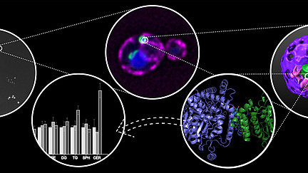
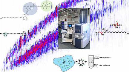
This central project expands the consortium’s technical capabilities in analyzing lipid function by developing protocols for a rapid and efficient isolation of cellular organelles, establishing workflows for mass spectrometry-based analyses of organellar lipidomes and proteomes, and generating a toolbox of functionalized lipids for monitoring intracellular lipid flows, capturing lipid effector proteins, and manipulating subcellular lipid pools.

A laboratory table with a large microscope, microsocopies can be seen on a screen.
© Lena Dehnen | Universität Osnabrück

Project Z2 - Dovile Januliene, Rainer Kurre & Katherina Psathaki : High-resolution imaging across spatiotemporal scales

This project provides a platform for advanced imaging across different spatial and temporal scales to resolve the highly dynamic organization of proteins and lipids within cellular membrane systems. It will establish a platform for high-resolution macromolecular structure determination and time-resolved cryo-EM, implement and further develop advanced light and electron microscopy approaches, and establish lipid imaging at the highest spatiotemporal resolution using novel lipid tools.

The logo of the Collaborative Research Center can be seen on a circle in the middle, from which dashed lines lead to smaller circles containing symbols related to biological research.
© Susanne Kunis | Universität Osnabrück

Project INF - Susanne Kunis, Michael Hensel & Arne Möller : Structured storage, metadata annotation, and analyses of experimental data

This project will further develop the current platforms for image data management and provide data management for additional data formats from proteomics and lipidomics mass spectrometry and flow cytometry. The project will interface the current data management platforms with an electronic lab notebook system for demands of SFB 1557 to enable the digitization of instrumental and experimental metadata alongside the data generation in experiments and analyses.

Career Support & Outreach

A group of about 30 people stand on a staircase and smile into the camera.
© Jan-Hannes Schäfer | Universität Osnabrück

Project MGK - Achim Paululat : Integrated Research Training Group

The IRTG complements the experimental training with training in a broader spectrum of subjects and provides opportunities for networking and personal development. The program includes regular lectures and progress reports, workshops on research methods, seminars on soft skills and networking opportunities through career seminars. All activities are embedded in a structured educational program with an individual development plan and a thesis advisory committee.

A collage shows a microscope, colorful pictures of a dividing cell, students filling out worksheets, and students holding signs.
© Chemiedidaktik u. Wissenschaftskommunikation, Zoologie | Universität Osnabrück

Project OR - Marco Beeken & Katia Cosentino : Outreach project of the SFB 1557

This project will accompany the SFB from an early stage with various innovative formats of science communication. The main aim is to present the central research questions of the SFB, and to explain how basic research in biology is linked to public interests. The project will promote the highly innovative infrastructural and technological portfolio offered by the SFB and the associated CellNanOs research building, which are flagships of the city and can be unique hubs for both research and public education.