Divisions and research groups

Colourful, microscopic image of tissue.
© A. Tomasso/ K. Bartscherer

Prof. Dr. Kerstin Bartscherer : Animal Physiology

Why can some animals simply regenerate injured tissue, while humans instead develop scars? The Animal Physiology division investigates this question – using modern 3D in vitro models and regenerating animals to unravel the secrets of regeneration and inspire new therapeutic approaches.

A collage of photos of mice, diagrams and a photo of Chadi Touma in a lab coat with a pipette in his hand.
© Behavioural Biology division

Prof. Dr. Chadi Touma : Behavioural Biology

The Behavioural Biology division researches molecular mechanisms of stress susceptibility, resilience and animal welfare. A wide range of animal species are investigated, from laboratory mice to farm animals to zoo and wild animals.

A collage of diagrams. The lettering ‘Fröhlich Lab’ at the top right.
© Bioanalytical Chemistry division

Prof. Dr. Florian Fröhlich : Bioanalytical Chemistry

The Bioanalytical Chemistry division investigates how cells regulate fat metabolism. Using baker's yeast as a model organism, the aim is to establish the molecular basis for understanding how disturbances in fat metabolism can cause disease.

A collage of an image of gel bands labelled ‘Protein purification of HOPS (six subunits)'; on the right are two 3-dimensional molecular structures, between which is the text ’3D model of HOPS (left) and CORVET (right)'.
© Structural Biology division

Prof. Dr. Christian Ungermann : Biochemistry

The Biochemistry division investigates cellular transport processes in the model organism Saccharomyces cerevisiae (baker’s and brewer’s yeast). The research focuses on the molecular mechanisms underlying the transport of proteins and organelles to the cell’s digestive compartment — the lysosome.

A hilly meadow with flowers, bordered by trees at the back.
© Thomas Fartmann

Prof. Dr. Thomas Fartmann : Biodiversity & Landscape Ecology

The Biodiversity and Landscape Ecology division investigates the effects of land use/climate change and disturbances on biodiversity. Forthermore, it focuses on monitoring species diversity and restoring habitats.

Against a green background you can see a photo of pigs, two hands holding soil with a plant, meat in a petri dish, a robin and insects that are obviously intended for consumption. ‘Willkommen!’ is written above it.
© Biology Didactics division

Prof. Dr. Susanne Menzel (Deputy: Dr. Florian Fiebelkorn) : Biology Didactics

Models of molecules, one is coloured in different colours, the others are grey.
© Biophysics division

Prof. Dr. Jacob Piehler : Biophysics

The Biophysics division studies structure, dynamics, and signaling of cytokine receptors in plasma membranes. Advanced fluorescence and single-molecule approaches are used to quantitatively analyze molecular mechanisms of cellular communication.

A collage of images of plants and microscope images.
© Botany division

Prof. Dr. Sabine Zachgo : Botany

The Botany division investigates the developmental processes and innovations of plants that have resulted in the enormous diversity of land plants and adaptations to variable living conditions.

A collage of various graphics and photos related to science; in the centre is a group photo of three women and one man smiling at the camera.
© Denisa Jamecna / Simone Reukauf Fotografie

Dr. Denisa Jamecna : Chemical Biology of Membranes

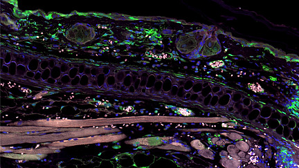
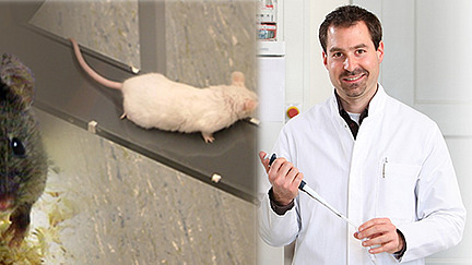
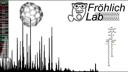
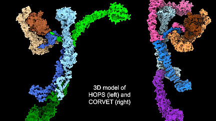
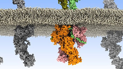
The Chemical Biology of Membranes research group investigates cellular lipid transport. Using synthetic lipid-like molecules, we study how lysosomes, cellular recycling organelles, distribute various lipids such as sterols and sphingolipids throughout membranes.

A collage of microscopy images and photos of diagrams and a pipetting process.
© Ecology division

Prof. Dr. Christian Kost : Ecology

A collage of microscope images, pictures of Petri dishes and a photo of bread with a filled wine glass and a filled beer glass.
© Genetics division

Prof. Dr. Jürgen Heinisch : Genetics

The Genetics division conducts basic research into stress response mechanisms and the associated signal transduction pathways in various yeasts and fungi as eukaryotic model organisms.

A collage of microscope images, including microorganisms, and diagrams.
© Microbiology division

Prof. Dr. Michael Hensel : Microbiology

The Microbiology division investigates molecular mechanisms of bacterial pathogenesis. Using the model organism Salmonella enterica, we unravel the pathogenic lifestyle in the host, host adaptation, and function of virulence factors.

Colourful microscopy images can be seen on the left and right, with schematic representations of cellular processes in the centre.
© Molecular Cell Biology division

Prof. Dr. Joost Holthuis : Molecular Cell Biology

The Molecular Cell Biology division investigates how cells assemble membranes from a structurally diverse repertoire of lipids, thereby creating the most advanced biomolecular systems in nature.

A colourful microscopic image
© RG Multicellular Signaling Dynamics

Dr. Lena Tveriakhina : Multicellular Signaling Dynamics

The Multicellular Signaling Dynamics research group investigates the dynamic properties of cell signaling pathways and how they control various cell fate decisions during the development and maintenance of multicellular organisms.

A collage of microscopic and graphic representations of nerve cells and cellular processes, as well as fingers holding tweezers.
© Neurobiology division

Prof. Dr. Roland Brandt : Neurobiology

The Neurobiology division investigates the development and degeneration of nerve cells at the molecular, cellular and systemic levels. A significant part of the work focuses on Alzheimer's disease, with the aim of developing the basis for new therapeutic approaches.

A man in a lab coat looks at an experimental set-up.
© Markus Scholz | WissenschaftsCampus Halle

Prof. Dr. Nico Dissmeyer : Plant Physiology

The Plant Physiology division investigates why some proteins remain in cells for a long time and can perform their functions, whereas other proteins can be very short-lived – and therefore only have a limited duration of action.

Graphical representation of molecules against a green background. At the bottom left, the word ‘Strubios’ is written in red.
© Structural Biology division

Prof. Dr. Arne Möller : Structural Biology

The Structural Biology division uses cryo-electron microscopy to determine the atomic structure of proteins and their molecular dynamics. It is particularly interested in transmembrane transporters, which are of great importance for the development of broad-spectrum resistance.

A graphic representation of ring-shaped molecules against a green background.
© RG Structural Biology of Photosynthetic Microorganisms

Dr. Dmitry Shvarev : Structural Biology of Photosynthetic Microorganisms

The Structural Biology of Photosynthetic Microorganisms research group investigates the molecular mechanisms of metabolism in phototrophic organisms – i.e. organisms that use light as a source of energy. Using cyanobacteria as a model and cryo-EM as the main method, the group researches chlorophyll biosynthesis, glucan degradation and transport processes.

A microscope image labelled ‘Nephrocytes’, ‘Heart Tube’ and “Muscles”; top left: ‘Zoology & Developmental Biology Lab (AG Paululat)’.
© Zoology division

Prof. Dr. Achim Paululat : Zoology

The Zoology division researches the genetic and molecular causes of congenital organ defects. The team is particularly interested in genes that ensure cell function. One area of research focuses on intracellular membrane systems and human heart disease, which are ‘recreated’ and analysed in fruit flies.